Search
Liens UNIL
Prof. Edward (Ted) Farmer
Our Research
We studied a mechanism which may underlie and maintain the high ratio of plant to animal biomass on earth. As a first step, we established that jasmonate, a small lipid derivative, mediates plant defense responses against herbivores. In a second phase of research, the laboratory used genetic approaches to study the rapid activation of these defense responses. The genes identified in that work shed light on both similarities and differences to electrical signalling mechanisms in metazoans.
Our past activities
IDENTIFICATION OF RICCA'S FACTORS
Ricca’s factors, first proposed in 1916 by Ubaldo Ricca, were thought to underlie electrical signalling in wounded plants. However, numerous attempts to define Ricca’s factors (e.g. by Umrath, Fitting, Schildknecht, Sibaoka, Pickard etc.) were unsuccessful.
We found that Ricca’s factors are proteins and that their transport in the vasculature underlies electrical signalling in Arabidopsis leaves distal to wounds. To our knowledge, organ-to-organ protein transport has not been implicated previously in electrical signalling in nature.
Long distance axial transport of proteins activates electrical signalling: https://www.cell.com/cell/pdf/S0092-8674(23)00108-3.pdf
MECHANOSENSATION IN LEAF VEINS
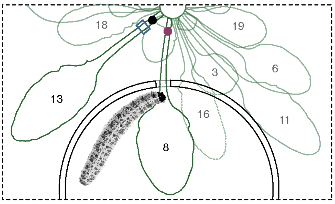
Epidermal cells on leaves, and in particular sensory trichomes, respond actively to non-damaging mechanostimulation. But do subepidermal cell populations respond to touch? We found that primary veins in leaves are touch-sensitive. Mechanostimulation triggers propagating electrical activity (as well as propagating Ca2+ transients) in these veins. Through genetic interventions we found that the phloem is the source of these signals. This work identifies a role for two P-type proton pumps in the remarkably high sensitivity of the phloem to environmental perturbations.
THE IMPORTANCE OF HAVING GOOD TEETH
We humans and most of the animals we cultivate need good teeth. The same is true of chewing insects. With each bite, herbivores activate the synthesis of the defense-inducing hormone jasmonate. Moreover, plants use multiple types of abrasive crystals and non-crystalline solids like silica to blunt the teeth and mandibles of the organisms that eat them. Why do they do this? There seem to be two reasons: Firstly, with bad teeth, the efficiency of digestion is reduced, and the growth of the herbivore is slowed. That’s the direct effect on the insect.
But there is more to the story. The smaller and the sharper the cut the less the plant responds, and herbivores frequently cut highly regular circles and semi-circles in leaves. This is mechanically economic (the insect doesn't have to move much) to do this. However, we found that some herbivores probably try to extract the maximum area of leaf tissue while leaving the minimal length of cut edge, thereby cleverly minimizing defense gene activation (Plant Cell 12, 707). To counter this, plants produce abrasive defense compounds like calcium oxalate. The sharp mandibles of chewing insects are blunted, and they now crush instead of cut. Crushing causes the release of jasmonate-inducing elicitors from plant tissues: more crushing, more defense elicitors.
We recently developed theoretical models for jasmonate synthesis activation by plant-derived elicitors of membrane depolarization. In these models, we show why tissue crushing favours plant defense activation and cutting doesn’t. In the models we envisage all plant cells to behave like stomatal guard cells. In essence, when cell membranes depolarize, the cells lose water. When cell membranes repolarize, they again take up water. This process, which depends on low water potentials cell walls, drives defense-inducing elicitor dispersal in wounded tissues. It’s exactly what the herbivore doesn’t want.
NEW: For more please read here: https://academic.oup.com/jxb/advance-article/doi/10.1093/jxb/erac449/6827807
RESISTING SUSTAINED ATTACK
The energetic cost of being bitten is high. The potentials of membranes that are depolarized in response to wounding need to be restored, and transient cytosolic Ca2+ increases need to be brought back to resting levels. Both processes require ATP-fuelled P-type pumps. We found that the Arabidopsis Ca2+ ATPase mutant aca10 aca12 failed to tolerate sustained insect attack. The insect-attacked mutant quickly lost its ability to produce leaf-to-leaf electrical signals and was consumed rapidly. Interestingly, ‘senescence-feeding’ insects kill the tissues they feed on. Based on our findings a good way to do this is to target Ca2+ ATPases which are expressed in the phloem. Curr. Biol. (2022) 32, 2517-2528. e6.
LONG DISTANCE WOUND SIGNALS
In response to wounding, jasmonates can be made within and proximal to wounds and also in distal tissues. Jasmonate synthesis can be activated by long distance wound signals other than mobile jasmonates. The nature of these signals has long been a mystery.
Showed that leaf-to-leaf wound signals that activate jasmonate synthesis move at speeds in the cm/min range (J. Biol. Chem. 283, 16400) and made the first constrained (minimum and maximum) velocity estimates for the speed of long distance wound signals that travel from leaf to leaf after wounding (J. Biol. Chem. 284, 34506). We showed that these long distance wound signals move through well-defined parastichies (J. Biol. Chem. 284, 34506 ; New Phytol. 197, 566).
Showed that clade 3 GLUTAMATE RECEPTOR-LIKE (GLR) proteins mediate electrical signalling in plants (Nature 500, 422).
Showed that the AHA1 proton pump functions to re-build membrane potentials after wounding (PNAS 116, 20226-20321).
We found that the first burst of jasmonate made in leaves distal to wounds in Arabidopsis originates from the activity of LOX6, an enzyme localized to xylem contact cells (New Phytol. 197, 566).
Developed aphids as living electrodes and used them to characterize GLR-dependent electrical signals in phloem sieve tubes (New Phytol. 203, 674).
In 2018 we discovered that two non-adjacent vascular cell types (xylem contact cells and phloem sieve elements) are necessary for the transmission of electrical signals from leaf to leaf (PNAS 115, 10178). Interestingly, endomembranes within these cells are implicated in controlling the ion fluxes underlying the electrical signals and this is consistent with other results from genetics studies (New Phytol. 216, 1161).
Using irregular xylem mutants we found that xylem cell wall integrity determined not only the architecture but also the velocity of slow wave potentials. These results (PNAS 116, 26066) are consistent with the xylem carrying elicitors of contact cell depolarization, an event we think underlies slow wave potential transmission.
In 2020 we published a model integrating the roles of both the xylem and phloem in slow wave potential progagation (New Phytol. 227:1037). Current research in the lab is based on challenging this model.
GLR GENES CONTROL TURGOR-DRIVEN LEAF MOVEMENTS
The sensitive plant Mimosa pudica displays striking leaf movements when touched or wounded. We found minute, damage-induced leaf movements in Arabidopsis thaliana that were similar to the large leaf movements seen in wounded M. pudica. Using genetic approaches we found that these turgor-driven leaf movements in Arabidopsis were dependent on the GLR3.3 and GLR3.6 genes. To our knowledge these are the first genes shown genetically to control damage-induced leaf movement : PNAS 116, 26066 (2019).
COORDINATING DEFENSE AND GROWTH: THE ART OF BONSAI
We developed the first assays that showed that wound-induced plant growth restriction was jasmonate-dependent (Plant Cell 19, 2470) and later found a mechanism that slows the growth of roots when shoots are wounded (PNAS 110, 15473). More recently, we have investigated the role of jasmonate in the art of bonsai (Plant Physiol. 184, 1172).
JASMONATES: DEFENDING CAPTURED CARBON
The approximately 500 gigatons of carbon in living terrestrial vegetation is potentially valuable food for animals, however, most living plant mass remains uneaten in nature. Our research aims to understand why this is and, specifically, how plants defend themselves. We discovered that a lipid-derived signal, jasmonate, controls antiherbivore defence responses and limits the activity of detritivores. The jasmonate pathway operates at a major nexus in the carbon cycle, limiting carbon flow from both living and dead plants to higher trophic levels. The laboratory uses genetic and electrophysiological techniques to study jasmonate pathway activation.
Proposed with C.A. Ryan a central role of jasmonate in the plant immune system (PNAS 1990, 87, 7713) and published the first model integrating wound signaling and damage-associated molecular pattern (DAMP) signalling leading to jasmonate synthesis and defense (Plant Cell 1992, 4, 129). The year 2020 marks 30 years of research on jasmonates and plant defense!
Performed the first experiments to investigate the effects of a signal transduction pathway on the feeding behavior of a vertebrate herbivore (Mol. Ecol. 21, 2534-2541).
We showed that wound-response leaf growth restriction is mediated by the jasmonate pathway (Plant Cell 19, 2470).
When leaves are wounded the aerial growth of plants is reduced. We showed how root growth in these plants is controlled genetically so that reduced growth of stems is balanced by reduced root growth (PNAS 110, 15473).
Developed the first assays to assess the contribution of jasmonate signalling to root (PLoS Gen. 11(6) :e1005300) growth restriction after wounding and developed novel shoot to root wounding assays for Arabidopsis seedlings (PNAS 110, 15473 ; PLoS Gen. 11(6) : e1005300).
We showed that the genetic architecture of the jasmonate pathway is different in roots and aerial tissues (PNAS 110, 15473) and identified cell-specific regulatory layers that prevent activation of the jasmonate pathway in healthy roots (PLoS Gen. 11(6) :e1005300).
Co-discovered (in parallel with the goups of J. Browse and R. Solano) members of the 'JAZ' family of repressor proteins and shown that a natural transcript from JAS1/JAZ10 plays a role in wound-induced growth inhbition. Also descibed the 'Jas' motif as a functional element in this protein (Plant Cell 19, 2470).
Discovered a new jasmonate (dnOPDA) and revealed a novel 'hexadecanoid' branch of the jasmonate biosynthetic pathway (PNAS 94, 10473).
Showed that monogalactosyldiacylglyerol (MGDG) is a likely in vivo substrate for jasmonate synthesis (PNAS 94, 10473).
Showed that cyclopentenone jasmonates (OPDA & dnOPDA) can act as signals (PNAS 98, 12837).
Described the principal roles of all four Arabidopsis 13-lipoxygenases in vegetative tissues : LOX2 makes the precusorsors of arabidopsides, LOX3 and 4 control wound-response growth restriction, LOX6 makes JA precursors in xylem contact cells (New Phytol. 197, 566 ; J. Biol. Chem. 284, 34506 ; Plant Physiol. 2020).
Quantitated (using microarrays and statistical analysis) the number of chewing insect-inducible genes that are regulated throught the jasmonate signal pathway (an estimated 67-84%) (Plant Cell 16, 3132) and shown that a specialist and a generalist insect activate this pathway almost equally.
Showed that jasmonate signalling promotes the establishment of an oxidising environment near a wound (Plant Physiol. 156, 1797).
We showed that endogenous jasmonate can move axially and radially from cell to cell in plants (Plant Physiol. 169, 2244).
Identified fou2, a wound-mimic mutant containing a single missense mutation in the vacuolar voltage-gated cation channel TPC1 (Plant J. 49, 889 ; Plant Cell Physiol. 48, 1775).
PHASE CHANGES ?
When wound-response electrical signals reach their destinations they trigger jasmonate synthesis rapidly; in a matter of seconds. The identification of a novel non-bilayer-forming galactolipid containing the jasmonate precursor OPDA suggested that membrane-depolarization-induced lipid phase changes in chloroplasts allow lipase action to initiate jasmonate accumulation (Morin et al., New Phytol. 240, 1484).
Oxylipins control a major biological transition: from detritivory to herbivory
Evolution from detritivory (feeding on dead tissues) to herbivory (feeding on living plants) is one of the major transitions in biology. We starved isopod crustaceans (detritivores) and let them loose on Arabidopsis and rice jasmonate synthesis mutants. The isopods started to feed on the living mutant plants but rarely damaged the WT. This shows that the absence of a signal pathway (the jasmonate pathway) in one organism causes a profound (transitional) effect on feeding in another (PNAS 106, 935).
MEMBRANES AS STRUCTURAL ANTIOXIDANTS
Tissues in which there is a high production of ROS (e.g. retina, neurons, thylakoids etc.) are particularly rich in oxidation-sensitive polyunsaturated fatty acids (PUFAs). At the subcellular level the membranes with the highest PUFA levels are often found where ROS are produced (e.g. mitochondria and chloroplasts).Why is this? Using genetic approaches we found that polyunsaturated fatty acids have roles as sinks for ROS. That is, membranes rich in these fatty acids can now be regarded as supramolecular antioxidants (J. Biol. Chem. 284, 1702; Ann. Rev. Plant Biol. 64, 429).
Showed that malondialdehyde (MDA) is an intermediate in a lipid repair cycle (J. Biol. Chem. 291, 13005).
Reactive Electrophile Species (RES)
Proposed that 'reactive electrophile species' (RES) could play roles in abiotic stress responses and in the 'abiotic component' of biotic attack in plants (Plant J. 24, 467; Nature 411, 854).
Demonstrated powerful biological activity in the ubiquitous and highly studied lipid peroxidation marker malondialdehyde (MDA) (Plant J. 37, 877) and showed that linolenic acid is the major source of damaging reactive molecules produced by non-enzymatic oxidation in leaves (J. Biol. Chem. 202, 35749).
Developed a whole-body malondialdehyde mapping technique for the in situ visualization of MDA in complex organisms (J. Biol. Chem. 202, 35749) and discovered inducible pools of MDA in stem cell-rich tissues including the pericycle (J. Biol. Chem. 287, 8954-8962).
Proposed the ‘lipid stress hypothesis’ with M.J. Mueller (Ann. Rev. Plant Biol. 64, 429). Stress triggers fatty acid de-esterification from membranes. Oxidized fatty acids induce chaperones that might act as binding surfaces for the displaced fatty acids that could otherwise disrupt hydrophobic protein-protein interaction surfaces.
Biologically active cell wall fragments
Showed that biologically active pectic fragments (oligogalacturonides) are egg-box conformers where oligogalacturonides are bound to calcium ions (J. Biol. Chem. 266, 3140).
Remorin: a new family of proteins
Discovered the remorin group of plasma membrane proteins (PNAS 86, 1539; Plant Cell 8, 2265). 'Remorins may be associated with the cytoskeleton or membrane skeleton i.e., in superstructures that help determine cell integrity and/or act as scaffolds for signaling in defense or development' (Plant Mol. Biol. 55, 579).
Missing phytoalexins from potato
Good candidates for low molecular mass antibiotics (phytoalexins) from the leaves of the historically and commercially important plant potato were unknown. They turned out to be relatively unstable molecules that had been described in vitro but that had previously escaped detection in living organisms. We identified divinyl ether fatty acids for the first time in nature in these leaves and proposed a role as antimicrobial compounds (Plant Cell 11, 485). They are the best candidates for the long-lost phytoalexins in potato leaves, the primary infection site for potato blight.
Community resources
Our laboratory has strongly supported the development of central, shared community resources in molecular biology and played a role in the establishment of the Lausanne Genomics Facility.